


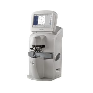
Canon Xephilio OCT-S1
Faster
100,000 A-scans per second combined with invisible 1,060 nm wavelength provide ultra-fast swept source technology maximizing data quantity of the patient’s eye while reducing acquisition time. Invisible scan lines ensure better patient collaboration and reduce the impact of patient eye movements.
Wider
With a single capture the swept source Xephilio OCT-S1 shows a large wide-field OCT image of up to 23×20 mm, which can be very beneficial for retina thickness observation of retinal detachment or retinitis pigmentosa. Mosaic imaging allows you to create an incredibly wide-field OCT image of approximately up to 31 x 27 mm.
Deeper
Canon’s deep scanning swept source technology allows better penetration of cataracts, hemor- rhages, blood vessels and sclera and at the same time optimizes capture of retinal and choroidal data – all in a single shot. With Xephilio OCT-S1 vitreous body and choroid appear in the same image with superior image quality providing more information for better patient care.
Xephilio OCT-S1
Wide-Field Sweft Source OCT
AI helps you save time and improve imaging
Canon’s Deep Learning technology Intelligent Denoise offers a new quality of OCTA images based on individual scans – without the need to acquire and merge multiple images. The revolutionary technology delivers images with greatly reduced image noise, increased detail and improved visibility within just seconds
Wide-field swept source imaging in one single capture
With Xephilio OCT-S1 Canon introduces revolutionary swept source technology allowing you to capture wide-field images of up to 23 mm in a single scan. Xephilio OCT-S1 enables superior penetration of dense objects and provides outstanding tomographic images. Intelligent Denoise, the system’s Deep Learning AI technology, offers a new quality of OCTA images in a single scan with greatly reduced noise, increased detail and improved visibility within just seconds.
Outstanding imaging is your best friend
Canon’s recognized optical expertise enables the Xephilio OCT-S1 to offer superb image quality with minimal scatter. The swept source technology results in enhanced penetration further into the deeper tissue structures such as the choroid and even the sclera. Imaging depths of up to 5.3 mm allows for detailed visualization of the vitreous body and choroid in a single scan while the high scanning speed of 100,000 A-scans/s reduces examination time and offers very high resolution scans.
Easy and quick operation
The Xephilio OCT-S1 utilizes a joystick for initial anterior alignment, but operation is also aided by several automated functions. It has built-in SLO for real-time retinal tracking and accurate follow-ups.
Visualize the microvasculature of the retina with OCT angiography
Non-invasive examination, results within seconds OCT Angio does not require fluorescein injection or pupil dilation, and the examination takes only seconds. SLO-based real-time tracking minimizes artefacts. Sophisticated image post-processing with 3D projection artefact removal enables excellent image quality. Angio Expert with freely selectable layers With OCT angiography even the smallest blood vessels can be observed in 2D and 3D. With Canon’s OCT Angio software, you can freely select layers to create the preferred image. Layers can be defined based on automatic segmentation or as a custom offset. OCT angiography is a sophisticated technology that detects the movement of red blood cells in the retinal vasculature and allows you to visualize tiny vessels in detail.
High density and single capture
The Xephilio OCT-S1 offers an enormous diversity of scan areas and scan densities for OCT Angiography examinations. While scan areas range from small (3 x 3 ~ 8 x 8 mm) to super large (23 x 20 mm), a high scan density of up to 928 x 807 pixels allows for visualization of small vessels at the same time.
Spesification
| Scan Speed | Max. 100,000 A scan/second | ||
| Horizontal Resolution | 30μm | ||
| Axial Resolution | 8μm | ||
| Light Source Wavelength | OCT: 1060nm, SLO: 780nm | ||
| Small Pupil Size | Φ3.0mm | ||
| Working Distance | 20mm | ||
| Retina Observation Method | Flying spot SLO | ||
| SLO FOV (H x V) | 23mm x 20mm | ||
| OCT Scan Width | 3~23mm | ||
| OCT Scan Depth | 5.3mm | ||
| Internal Fixation | “x” shape display on retina: green | ||
| External Fixation Light | EL-1 (option) | ||
| Dimension and Weight | Dimension (W x D x H) | 510mm x 330mm x 590mm | |
| Weight | 35kg (main unit only) | ||
| OCT Scan Parameters | Retina scan mode | Custom 3D | 512 x 512 1024 x 128 1024 x 1024 2048 x 512 Vertical/Horizontal |
| Multi-cross | 1024 x 1024 2048 x 2048 4096 x 4096 Averaging: 1–50 |
||
| Cross | 1024 x 1024 2048 x 2048 4096 x 4096 Averaging: 1–200 |
||
| Radial | 1024 2048 4096 12 directions (15 degree intervals) Averaging: 1–50 |
||
| OCTA | Small: 232 x 232 (3 x 3 ~ 8 x 8mm) Medium: 464 x 464 (4 x 4 ~20 x 20mm) Large: 696 x 696 (6 x 6 ~ 20 x 20) Super Large: 928 x 807 (23 x 20mm) |
||
Reviews
There are no reviews yet.